Microbubble technology is highly suited for studies in cancer research. MB arrays are molded in polydimethylsiloxane (PDMS) and comprise an ideal platform to sort tumor initiating cells (TIC, cancer stem cells) and study their fate and drug resistance.
Advantages of MBAtm for cancer research
- PDMS is optically clear with an elastic modulus (2 MPa) which is more similar to soft tissues (e.g. skin = 0.85 MPa) than hard tissue culture polystyrene (2500 MPa).
- PDMS is hydrophobic which favors anchorage independent cell growth (anoikis resistance), a characteristic phenotype of TICs.
- The unique spherical architecture and nanoliter MB volume concentrates cell secreted factors allowing rapid conditioning of the microenvironment , promotes single cell clonal proliferation.
- Clonal proliferation of nonadherent aggregates (tumor spheroids) increases expression of stem cell markers, migratory/invasive characteristics, and resistance to chemotherapeutics.
example APPLICATIONS
The MB microenvironment has been exploited to study the epithelial to mesenchymal transition (Chandrasekaran et al., 2011) which is important process in cancer metastasis.
MB arrays have been exploited to quantify the clonogenic potential of cancer stem cells (Chandrasekaran and DeLouise, 2011) and the metastatic potential of melanoma cells (Chandrasekaran et al., 2016).
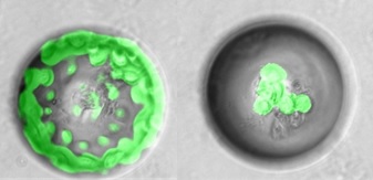
exhibit morphologic heterogeneity
The heterogeneity of tumorigenic squamous cell carcinoma (SCC) cells was dissected at the single cell level to discover morphologically distinct cell colonies and rare drug resistant cells (Pu et al., 2017)
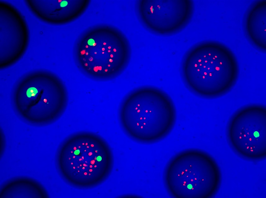
green – live cells, red – dead cells
MB arrays have been used to study the drug resistance of multi-cellular tumor spheroids in a 3D perfusion culture system (Agastin et al., 2011) and to investigate the effect of homo and heterotypic interactions on cancer cell function (Chandrasekaran et al., 2012).